


Where Does Ionizing Radiation Come From, and How Does It Impact You?
Ionizing radiation is a topic that covers a lot of ground, and whether you are interested in obtaining a radiological test or you are simply worried about radon gas, there is a lot that you can find out about it. In the following paragraphs, we are going to go over some of the most frequent sources of ionizing radiation as well as the effects that it may have on you. We are going to discuss the risks associated with radioactive radon, the risks associated with neutrons in emergency situations, and the risks associated with medical diagnostic tests.
Ionizing radiation is capable of exhibiting a variety of features, including the following:
- Radiation with high frequencies and energy is known as ionizing radiation. Examples of this kind of radiation include X-rays, gamma rays, and certain forms of UV radiation.
- Ionizing radiation has the capacity to ionize atoms, which means it has the power to cause atoms to either acquire or lose electrons, which ultimately results in the production of ions.
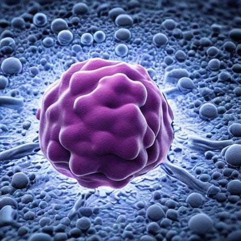
- Damage to living tissue and DNA may be caused by ionizing radiation, which can then result in adverse health consequences such as cancer and genetic abnormalities.
- It is possible for ionizing radiation to penetrate a wide variety of materials, including solid objects and the human body.
- Ionizing radiation may be detrimental to living beings; thus, it is imperative that preventive measures, such as shielding, be implemented to reduce the amount of exposure that these species get.
-
Ionizing radiation may be put to a number of useful applications, such as imaging in the medical field, the treatment of cancer, and the disinfection of medical equipment.
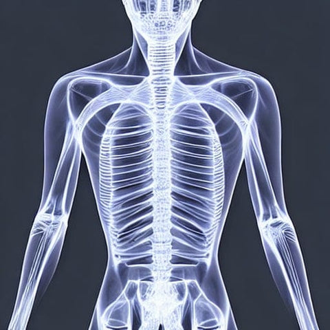
X-rays and Other Forms of Radiation From Diagnostic Medical Procedures
Radiation is often used in a significant capacity throughout the diagnostic testing process. In addition to assisting medical professionals in the diagnosis and treatment of patients, it also has the potential to bring on a variety of undesirable side effects. X-rays, in example, are an instrument that is often used in the diagnosis and treatment of a wide variety of conditions, including fractured bones. They are also capable of identifying the existence of life-threatening disorders such as cancer and other conditions. However, if they are used for an extended period of time, they may cause injury. For instance, new research has shown a correlation between exposure to radiation and an increased likelihood of acquiring cancer later in one's life.
Ionizing radiation is known to be hazardous to human health, however the amounts that are often encountered are rather low. In point of fact, the quantity of radiation that a person gets from a diagnostic test is often far less than the amount of radiation that the typical person receives from the natural background radiation in their house. As a consequence of this, medical x-rays have become the single most important source of exposure to radiation produced by human activity for the majority of individuals in the United States.
During a medical imaging examination, the quantity of radiation that is exposed to a patient is often measured in terms of the effective dose. This value is expressed as a millisievert (mSv), which is equal to 1/1000th of a rem. It is typically determined by taking into account the size of the subject as well as the imaging technique that is being carried out. Even while a chest CT exposes the patient to a greater dose of radiation than a standard x-ray, this dose is still several orders of magnitude lower than the amount of radiation that may be received from being exposed to a nuclear explosion.
Radiation is used in a variety of diagnostic procedures, such as mammography and positron emission tomography, to assist medical professionals in determining a patient's overall state of health. These kinds of diagnostic examinations are often regarded as the method that provides the clearest view inside of a body. Some of these examinations don't hurt, yet they nevertheless provide you valuable information. It is crucial to discuss your exposure with your doctor if you are going to be through a regular series of x-rays. You should also inquire about the advantages and hazards associated with having a series of x-rays done.
Other diagnostic procedures, such as computed tomography (CT) and nuclear medicine imaging, are further examples of diagnostic procedures that make use of radiation. Although some of these tests are risk-free, others entail very high dosages of the substance being tested for. Even CT brain perfusion scans have been linked to unsafe levels of radiation exposure, according to some investigations.
Imaging procedures used in medicine may expose patients to radiation, but there are several precautions that can be taken to reduce this risk. One option is to limit diagnostic procedures to those that are particularly relevant to the illness that is under investigation. Another thing you should do is record all of your test findings. You may guarantee that you are receiving the appropriate test at the appropriate dosage by maintaining open lines of communication with both your treating physician and the imaging staff.
If you want to protect yourself against the risks of ionizing radiation, the best thing you can do is limit the kind of diagnostic medical tests you get to those that are strictly necessary for treating your disease. In addition, do not fail to mention your pregnancy to your primary care physician.
The radiation emitted by radon gas
Radon is a radioactive gas that may be found collecting in the soil, rock, and water that is found below structures. Alpha and beta rays are emitted by the decay products of this element. They contribute to the development of lung cancer by being deposited in the respiratory system. Ionization is caused in a significant way by the radioactive particles. Because of this, being exposed to radon may raise one's chance of developing lung cancer higher. Smokers are at a larger risk than non-smokers.
Radon is referred to be a "confirmed human carcinogen" due to the fact that it has been shown to cause lung cancer in humans. It is believed that radon is responsible for between 3% and 14% of all fatalities caused by lung cancer. Radon has the potential to cause DNA damage in cells if it is not removed from the environment or treated.
At the beginning of the 20th century, it was common practice to subject patients to radon therapy for therapeutic reasons. Researchers found that cancer cells were susceptible to being killed by radioactive particles found in the gas. In order to investigate the impact that radon has on living creatures, a number of different experiments were carried out. During one of the experiments, a set of CR39 SSND detectors was used in order to collect data. Both were positioned around 20 centimeters away from the wall and observed for a period of three months. These detectors were calibrated to determine the levels of radon and thoron in the air.
The way the structure was constructed had an impact on the amount of air that was exchanged while the test was being conducted. According to the findings, radon was most prevalent in dwellings that included substantial ventilation. Radon did not get less concentrated as rapidly indoors as it did outside in the environment. An equilibrium factor was reached when the amount of radon in the air was equal to the rate at which air was being exchanged.
Radon has a propensity to build up in confined environments, which is another contributor to the elevated levels of the gas seen inside. They may enter a structure by any number of entry points, including windows, openings around pipes, or fissures in the floor. Radon concentrations are not uniform throughout the country due to the fact that the rate of air exchange is directly proportional to the building's structure. For instance, a broad stretch of coastline may have a radon concentration of 0.1 becquerels per cubic meter.
Radon is a radioactive gas that has an ionizing impact and may lead to a variety of lung illnesses. More specifically, it may cause harm to the epithelial cells that line the bronchi. Radon exposure raises the risk of developing lung cancer, which is already 10 times more likely among smokers than in those who have never smoked. Additionally, the additive impact of radon plus smoking contributes to an increased likelihood of developing lung cancer.
Uranium is the primary component of the radioactive gas known as radon. Uranium is a naturally occurring element that may be found in rocks, soil, and water. Radon is produced when uranium undergoes the decay process. Radiation of the alpha, beta, and gamma varieties is emitted when the parent atom breaks down into uranium-238 and 214Rn. Radon is a radioactive gas that may cause lung cancer, and alpha particles have been linked to this disease.
The United States Environmental Protection Agency (EPA) advises that those with radon exposures below 4 pCi/L should address the issue since the risk of radon-induced lung cancer grows with increasing radon exposure. Radon exposure is measured in picocuries per liter. The International Atomic Energy Agency (IAEA) publishes safety guidelines to assist national governments in the process of formulating radon action plans.
Radon was identified by the World Health Organization (WHO) as the second biggest cause of lung cancer in 2010. Smoking was shown to be the primary cause of lung cancer. The World Health Organization (WHO) keeps an eye on radon policy and country radon standards.
Emergency Radiological Situations That Include Neutron Radiation
In the event of a radiological emergency, neutron radiation poses a significant threat to human health and requires rapid response in order to reduce the likelihood of suffering an injury. The dangers are significant, and they might lead to life-threatening injuries or even death. Because of these factors, those who work in health care must be knowledgeable with the concepts of radiation safety. In addition, all of the staff are required to wear dosimeters so that they can monitor their exposure levels.
The most common way to get infected is via the skin, and this often takes the shape of a cut or other kind of wound. However, there is also the possibility of contamination on the inside. This may occur either by the inhalation of radioactive particles or the consumption of a radioactive material. Decontaminating the wound and removing any metallic shards that may be present is essential in the event that the wound is exposed to the outside environment. Decontamination and removal of any exposed metal may be accomplished with the use of forceps and tongs.
Radioactive substances may be found in the environment in the form of cosmic rays that come from the sky as well as in the form of materials that have been created by humans. These chemicals have the potential to inflict significant harm on the body and perhaps result in cancer. Because of this, emergency centers need to be ready to treat huge numbers of patients who are unwell and potentially polluted.
An emergency situation involving radiation may be caused by a number of different events, including an accident, the deliberate release of nuclear material, or the deployment of a radiation exposure device in an area with a high population density. In order to effectively react to catastrophic crises, medical physicists and emergency medicine professionals need to be well-prepared. Additionally, hospitals need to be ready to take patients whose bodies have been tainted by radioactive substances and should be prepared to do so.
A radiologic examination is performed on a patient before they ever enter a hospital to be treated. A Geiger-Mueller probe is used for this procedure in order to determine whether or not radionuclides are present. In the event that a person has been exposed, the removal of the radioactive materials requires either a punch biopsy or another technique. Documenting the results on an anatomic chart as soon as possible is of the utmost importance.
Burns to the skin and nausea are the acute radiation syndrome symptoms that are most often seen. The higher the dosage of radiation, the more severe these symptoms become. Diarrhea and vomiting are two symptoms that may be brought on by emotional stress and worry brought on by the perceived risk of radiation.
Thankfully, the negative effects that radioactive particles have on living organisms may be mitigated or eliminated entirely with the help of a control software. Being well-prepared and knowledgeable about the dangers posed by radiation is the most efficient method for mitigating the effects of potential radiation emergencies. Having an awareness of the concepts of radiation safety may assist emergency responders evaluate the risk and take appropriate precautions.
In addition, arrangements should be taken to guarantee that people can rotate in order to lessen the likelihood of being exposed to the virus. It should not be difficult to get any of the necessary materials and equipment. In addition to that, there need to be a strategy for treating those who have been polluted.
Emergencies involving radioactivity are often catastrophic and have the potential to have a significant influence on both public and economic health. They are also capable of causing a wide variety of diseases and damage. Radiation is not a naturally occurring material, yet it has the potential to inflict considerable psychological damage.
Looking for ways to stay healthy? View our selection of emf protection products.


Leave a comment